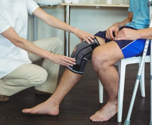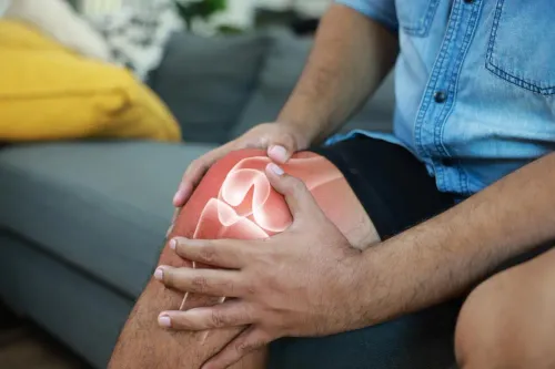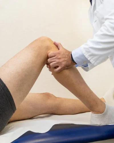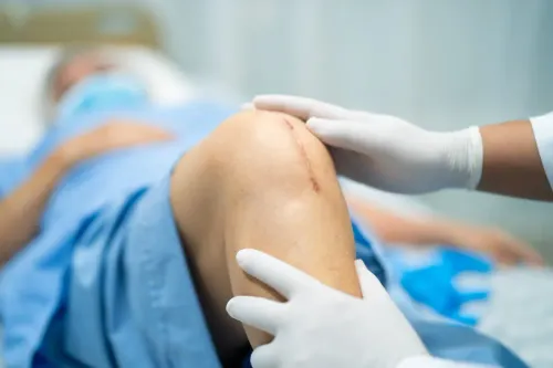
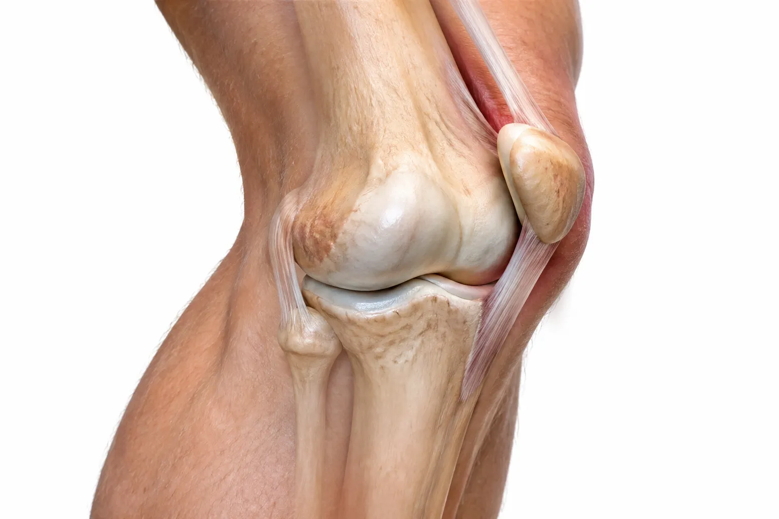
A patellar dislocation occurs when the kneecap moves completely out of its normal position, usually toward the outer side of the knee. It can happen during sport, after a sudden twisting movement, following an awkward landing, or because of direct impact.
For some patients, the kneecap moves back into position spontaneously. That can create the impression that the problem has resolved. However, the structures responsible for stabilising the kneecap may have been injured, making proper assessment important even after the visible deformity disappears.
What Does a Patellar Dislocation Feel Like?
During the injury, patients may experience a sudden shift or pop followed by significant pain. The knee may appear deformed if the kneecap remains displaced.
Other symptoms can include:
- Rapid swelling
- Difficulty straightening the knee
- Tenderness around the kneecap
- A feeling of instability
- Bruising
- Difficulty bearing weight
- Fear that the kneecap will slip again
After the kneecap returns to position, pain and swelling may persist.
Why Imaging May Be Recommended
A patellar dislocation can affect more than the stabilising ligament.
The event may injure cartilage or cause a small piece of cartilage and underlying bone to become detached. X-rays can help identify fractures and assess knee anatomy, while MRI may provide information about ligament injury, cartilage damage, bone bruising, and associated abnormalities.
Imaging decisions depend on the clinical findings rather than being identical for every patient.
First Dislocation Does Not Automatically Mean Surgery
Many first-time dislocations can be managed without an operation, particularly when there is no major loose fragment or other indication for surgical treatment.
Initial care may include appropriate support, swelling control, gradual restoration of movement, and physiotherapy.
Rehabilitation commonly focuses on restoring quadriceps function, hip strength, balance, movement control, and confidence.
The objective is to help the kneecap track appropriately while preparing the patient to return to normal activity.
When Repeated Episodes Become Important
A second or third dislocation changes the clinical picture.
Repeated instability can indicate that the medial stabilising structures are no longer providing adequate control. Certain anatomical features may also increase the likelihood of recurrence.
Specialist evaluation becomes particularly useful when a patient experiences:
- Multiple dislocations
- Frequent partial slipping episodes
- Persistent apprehension
- Ongoing swelling
- Mechanical catching
- Difficulty returning to sport
- Significant cartilage injury
Repeated instability should not simply be accepted as an unavoidable consequence of the first injury.
Returning to Sport Requires More Than Pain Relief
An athlete may feel substantially better before strength, balance, landing mechanics, and confidence have fully recovered.
A structured return-to-sport process can evaluate whether the knee is ready for running, cutting, jumping, pivoting, and sport-specific demands.
This is especially relevant for active patients across Mumbai, where football, cricket, running, gym training, racquet sports, and recreational fitness place different loads on the knee.
Dr Pranjal Kodkani's sports orthopaedic practice assesses patellar instability with attention to ligament injury, cartilage health, knee anatomy, and the patient's activity goals.
A patellar dislocation is therefore not simply a kneecap that temporarily moved out of place. Understanding why it occurred and whether the knee remains unstable can help guide safer rehabilitation while identifying patients who may need further treatment.
Keywords